MATRIX applications
All types of materials (cement, filtration membranes, batteries, etc.), biological samples (organisms, organs, plants, cells, etc.) and natural samples (soils, sediments, meteorites, etc.) can be characterised using a correlative and multi-scale approach. The issues addressed are very broad, particularly in relation to the environmental and energy transition, archaeology and new materials.
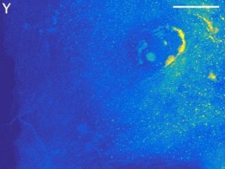
Studying the transfer of metal contaminants from soil to plants
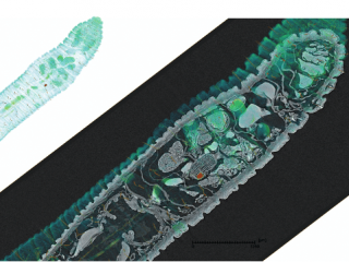
Example. Location of yttrium (Y, a rare earth) in alpine plants Saxifraga paniculataknown for its high tolerance and interesting potential for metal accumulation. LA-ICP-MS mass spectrometry and synchrotron X-ray fluorescence spectrometry (micro-XRF) were used to locate yttrium (Y) in plant tissues (roots and leaves) and identify co-localised elements.
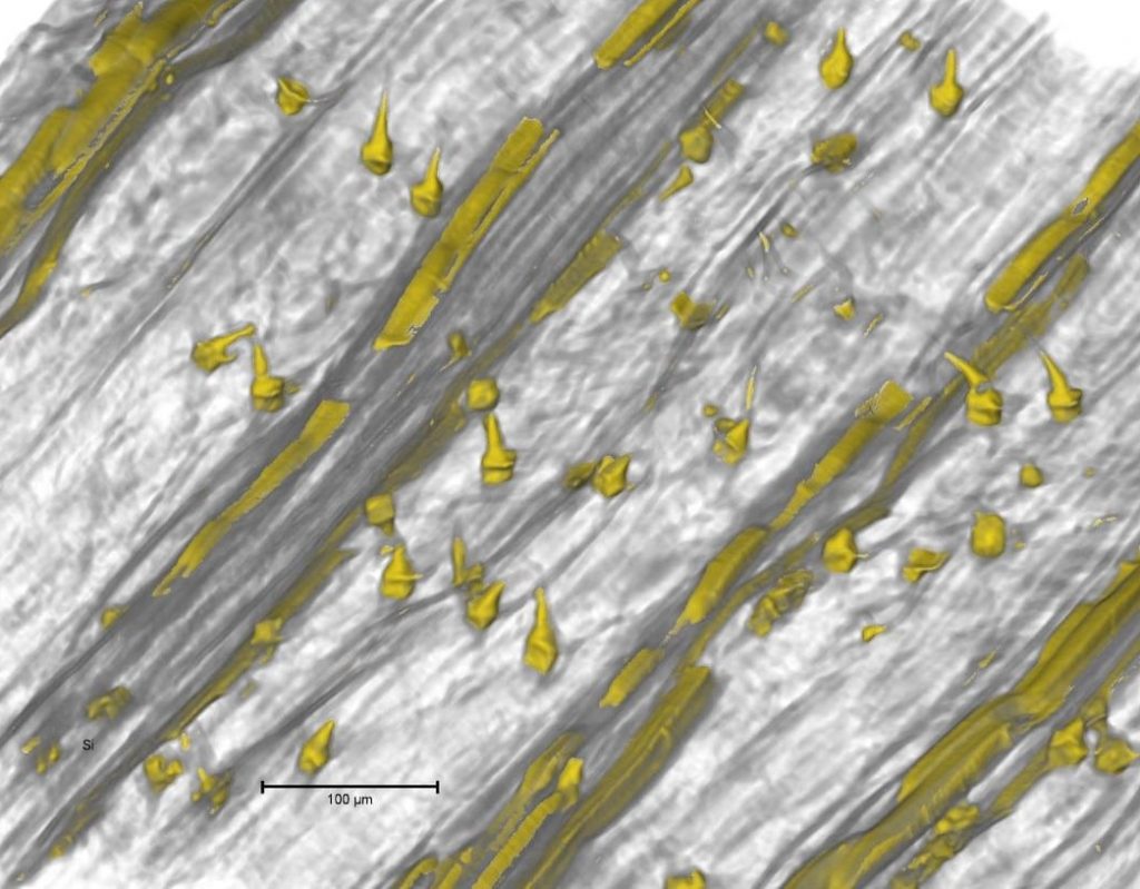
Observing plant defence mechanisms
Ex. In-situ counting of phytoliths in durum wheat leaves under increasing water stress. In 3D images of wheat leaves (micro-CT), silicified trichomes and elongated phytoliths can be seen, which have the role of strengthening the leaf structure. In the presence of silicon, the leaves accumulate phytoliths in their veins, which improves the overall development of the plant.
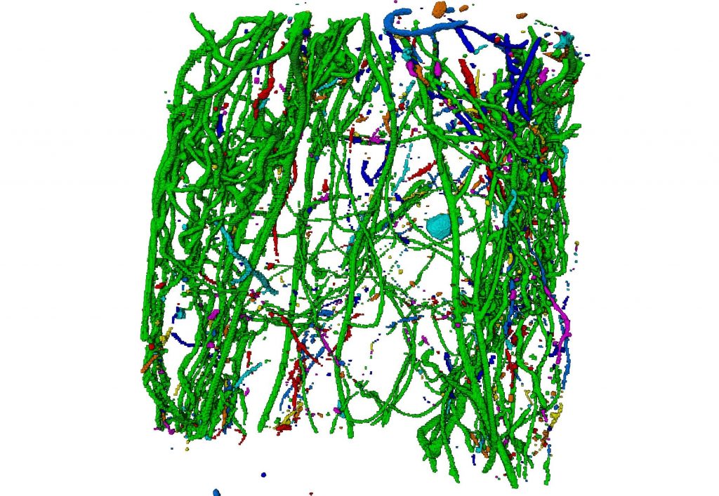
Analyse the architecture of a root system
E.g. Assessment of the toxic effects of a soil naturally rich in metals by monitoring the development of the root system of fescue plants (Festuca laevigata). Micro-CT enables 3D analysis of the architecture of the root system, including the finest roots (a few tens of µm in diameter).

Assessing the environmental impact of waste reuse
E.g. Monitoring the behaviour of Cu and Zn in a tropical soil-water-plant system following the spreading of pig manure. LAnalysis micro-XRF of pig manure and the statistical processing spectra obtained by SIMPLISMA can be used to identify the co-location of Cu and Zn with the other elements in the liser, and then to formulate hypotheses about the nature of their carrier phases and their speciation. Metal speciation can then be validated using synchrotron X-ray absorption spectroscopy (XAS).
Characterise a material, soil or sediment
Identification of the nature of the crystallised phases, minerals and/or clays present (XRD), detection of metal contamination (micro-XRF), quantification of porosity (micro-CT and nano-CT) ...
Cornu et al, Geoderma, 2022. Allophanes, a significant soil pool of silicon for plants.
Fine-tuning the porosity of new materials
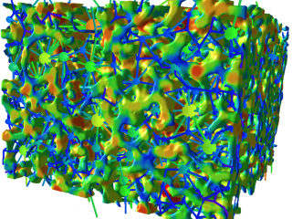
E.g. Characterisation of a silica monolith synthesised for the treatment of water contaminated by pharmaceutical products. The morphology of its pore network (macropores) is characterised by micro-CT: number, size, connectivity, tortuosity, etc. of the pores can be quantified.

Tracking variations in the internal structure of materials
E.g. Monitoring the swelling of the leaves of a lithium battery (Li-ions) during its charge/discharge cycles. The 3D image of the entire battery (low-resolution micro-CT, 1 vx = 42 µm) is used to reposition on the same area between two states of charge (SOC) for high-resolution micro-CT acquisition (1 vx = 1.6 µm). Analysis of the 3D images provides a measure of the thickness of the electrode and enables its swelling to be quantified.
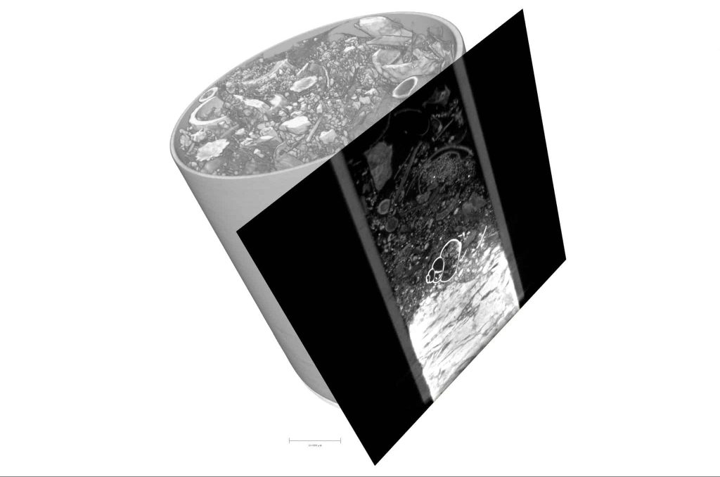
Locating nanoparticle aggregates in complex matrices
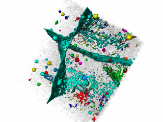
Ex. Detection ex-vivo and location of nanomaterials (CeO2-NMs) in mouse lungs. The correlative, multi-scale approach used (combining micro-CT, nano-CT, micro-XRF, XAS and histological observations) enabled us to visualise the bio-distribution of CeO2-NMs at the scale of an entire lobe and at the cellular level, i.e. in macrophages.
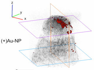
Ex. Localisation of gold nanoparticles (Au-NPs, 12 nm) at the apex ofArabidopsis Thaliana. The combination of 2D imaging (dark-field microscopy combined with hyperspectral, DF-HSI) and 3D imaging (X-ray nano-tomography) is improving our ability to detect and visualise nanoparticles in plant tissues, right down to the cellular level. The processes that control the uptake and distribution of Au-NPs in plant tissues can be identified (e.g. accumulation of mucilage that traps Au-NPs).
E.g. Determination of the accumulation, localisation and speciation of silver nanoparticles (Ag-NPs) in earthworms following the use of nanopesticides. The correlative approach used to assess accumulation capacities and mechanisms physiological factors involved in detoxification in Ag.

Tracking particle transport in a crack
For example, guaranteeing the safety of a radioactive waste package in an accident scenario where its mortar containment barrier cracks. Micro-CT allows in-situ visualization of model aerosol particles that have migrated into the crack. The particle transport mechanisms (retention and/or diffusion) involved within the cracked mortar can be identified.
Assessing the durability of a material
Characterisation of the deterioration of materials (paint, cement, coatings, plastic, glass, etc.) during the various stages of their life cycle (use, end of life, etc.).
E.g. Characterisation of the alteration profile in the surface zone of a cement subjected to leaching. The chemical, mineralogical and structural (porosity) evolution can be quantified using an approach combining micro-XRF, micro-DRX, micro-CT and nano-CT.
E.g. Monitoring the appearance of cracks (micro-CT) in a construction material subjected to freeze/thaw cycles.
Performing a non-destructive test
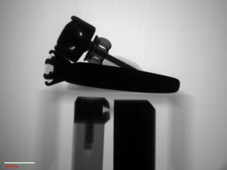
E.g. Rapid detection of defects in electronic components or solder joints by recording 2D X-rays (micro-CT).
Example. Checking the quality of a sun cream. 2D X-rays with very high spatial resolution (nano-CT) provide information on the optimal dispersion of nano-TiO2 for better UV absorption efficiency.
Create 3D model libraries
Micro-CT digitisation of a large number of objects to create 3D collections for the general public, education or research.
Example. Animation and integration of 3D models of arthropod specimens (ants) in a virtual reality environment to raise public awareness of the neglected biodiversity of French Guiana (eBREVE project).
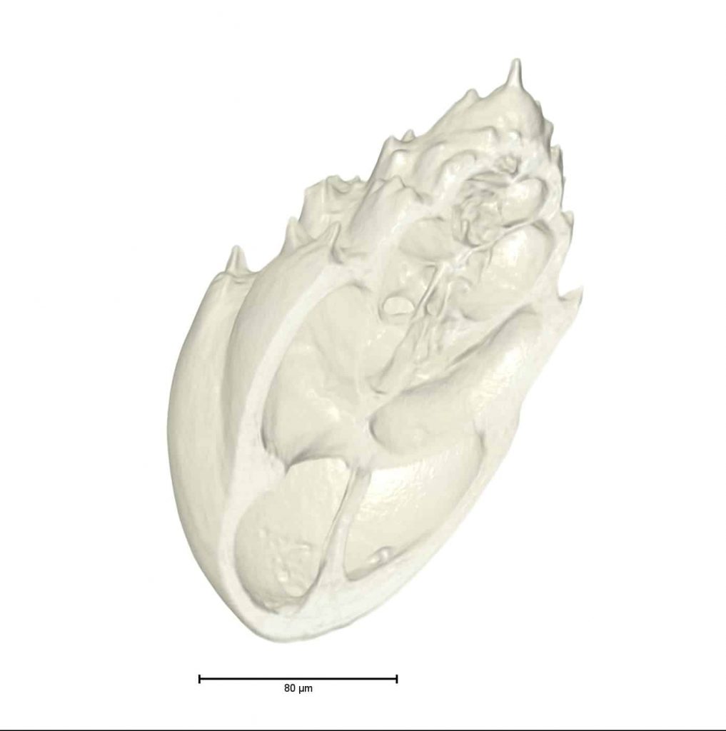
E.g. Creation of a library of micropalaeontological objects (foraminifera) to extend the possibility of observing and describing educational material in 3D beyond practical sessions (MicrovirtualPal project).
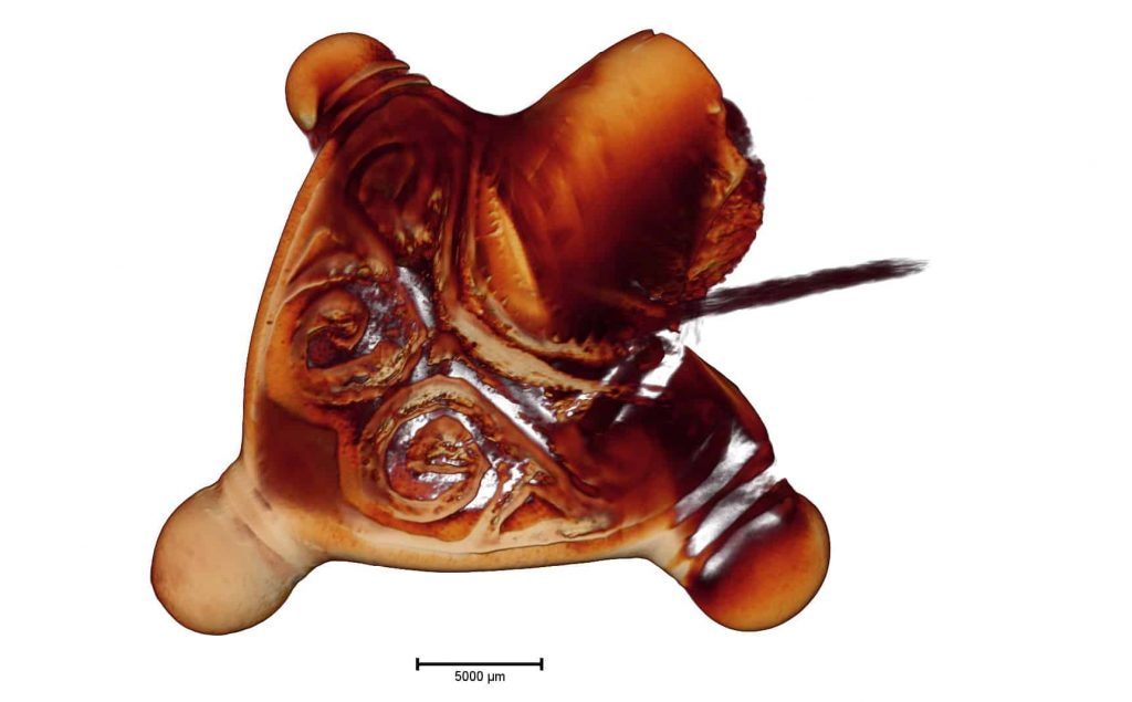
E.g. Creation of a 3D ichthyological database to help with the osteological identification of small bones of present-day and archaeological teleosts (bony fish) (Ictyo3D project).
Helping to identify archaeological objects
Non-destructive X-ray techniques are the techniques of choice for analysing archaeological objects!
E.g. Optimising the restoration of archaeological objects after determining their chemical composition (e.g. type of metal alloy) using micro-XRF.
E.g. Creation of ceramic groups from a chemical database constructed using portable X-ray fluorescence (pXRF) analysis.
E.g. Virtual manipulation and analysis of very fragile archaeological objects after 3D micro-CT scanning.
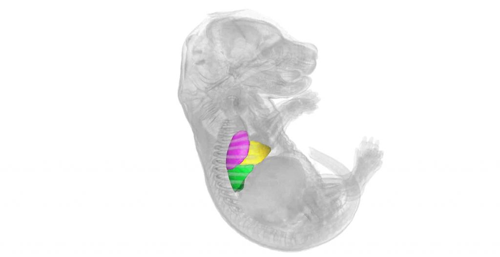
Monitoring embryonic development
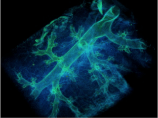
Ex. Verification of the correct development of the pulmonary system of a mouse embryo. 3D imaging (micro-CT) eliminates the delicate and time-consuming step of dissecting and preparing serial sections.
A micro-CT image (volume 1x1x1 mm31 vx = 1 µm) corresponds to the observation of 1000 histological sections 1 µm thick!