MATRIX Tools
The particularity of the platform lies both in the complementarity of the equipment available and in its exceptional performance in terms of spatial resolution. All types of materials can be imaged in 2 and 3 dimensions with a spatial resolution ranging from several hundred micrometres to about ten nanometres.
MatriX's instruments include an X-ray diffractometer, a number of X-ray fluorescence spectrometers, including a new-generation microscope funded by the European Commission.EquipEx+ Imagine2two high spatial resolution X-ray tomographs (micro and nano X-ray tomographs) acquired as part of theEquipEx NANO-IDIt also has a sample preparation laboratory and a computer system dedicated to data processing and image analysis.


X-ray Diffractometer
X-ray Diffractometer (X'Pert Pro MPD, Panalytical) to carry out analyses on powder (DRX) and spatially resolved micro-analyses using a 100 µm diameter capillary (micro-DRX).
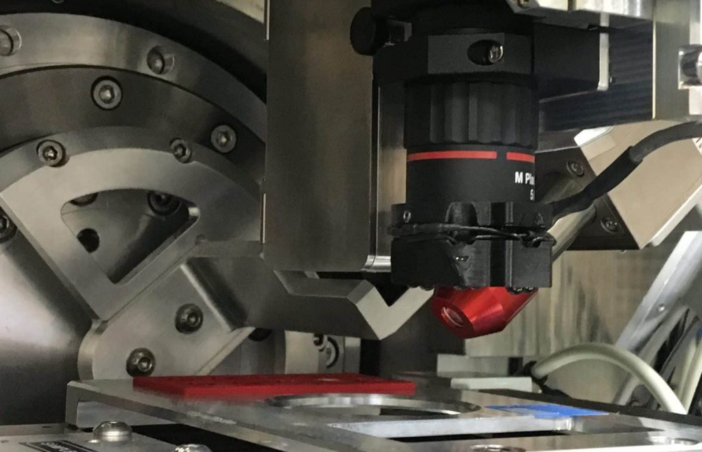
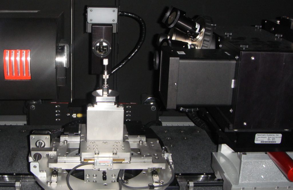
New-generation X-ray analytical microscope
Innovative device (AttoMap-310, Sigray), acquired as part of theEquipeX+ Imagine2offering the highest spatial resolution (down to 7 µm) and sensitivity (down to ppm) available in the laboratory, for rapid chemical mapping of the distribution of elements: from trace metals to light elements down to carbon (<1%).
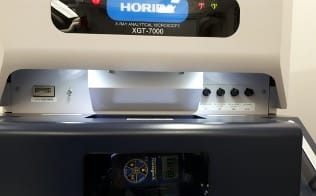
X-ray analytical microscope
Device (XGT-7000, Horiba) coupling micro X-ray fluorescence (micro-XRF) with X-ray microscopy to perform chemical micro-analysis and 2D chemical imaging (hyperspectral mapping) with a spatial resolution of 10 and 100 µm.
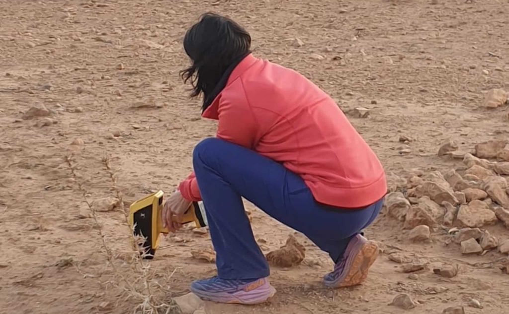
Portable XRF machine
Portable device (X-200, SciAps) to carry out qualitative and quantitative elemental analyses on site and/or on large samples.
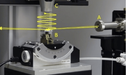
HERMES-TXRF Prototype
Prototype developed in collaboration with the FAME of the ESRF.
The prototype of a wavelength-dispersive microfluorescence X-ray spectrometer (HERMES for High Energy Resolution Microscope for Environmental Sciences) allows X-ray fluorescence analyses with a very high energy resolution, of the order of 1-10 eV. Its objective is to improve the sensitivity to light and trace elements and to access in-situ the oxidation states of certain metals.
The prototype of a total reflection X-ray fluorescence (TXRF) spectrometer aims to perform rapid and quantitative elemental analysis on powders. The advantages of such a set-up compared to conventional XRF are multiple: increased sensitivity and improved quantification of elements.
RX micro-tomograph
Device (MicroXCT-400, Zeiss) (acquired as part of theEquipeX NANO-ID) for multi-scale 3D imaging using X-ray absorption contrast, with spatial resolution ranging from 0.7 µm to 65 µm (micro-CT).
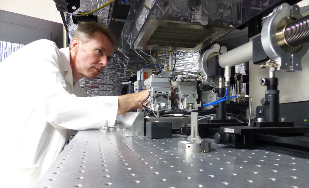
Live lab: visit our equipment!
Nano X-ray tomograph
Device (UltraXRM-L200, Zeiss), the only one of its kind in France (acquired as part of theEquipeX NANO-ID), for 3D imaging at very high spatial resolution (150 and 50 nm) in absorption contrast and phase contrast (nano-CT).

Sample preparation laboratory
Equipped laboratory:
- an optical stereo microscope for handling and fixing small samples (Leica M80)
- a Zeiss microscope adapted to the deposition of markers (micrometric gold beads).
- of a supercritical drying apparatus, bypassing the supercritical point of CO2 (EM CPD 3000, Leica Microsystems).
- a precision micro-saw (IsoMET 4000, Buehler) for the production of thin sections (minimum thickness of 200 µm)
- a micro-machining device (MINITECH CNC Mini-Mill/GX)
- a 3D printer (RAISE 3D PRO2) for designing specific sample holders
- equipment for sample inclusion and polishing
- small equipment dedicated to the preparation of samples for XRD analysis (grinding, phase separation, extractions, chemical treatments).

Image analysis
A data processing room for the Matrix platform instruments has been specially fitted out. It provides access to 3 workstations equipped with:
- Licences AVIZO and OSR Dragonfly for 3D image processing and analysis
- Open-source software (ImageJ, iMorph, PyMCA) for 2D and 3D image analysis
- Mineralogical database for interpreting X-ray diffractograms.
The storage capacity of the hard disks and a storage server (30 TB dedicated to X-ray tomography) allow data to be kept for at least one year.